
[ad_1]
Karly Cohen
Photographer
Karly Cohen
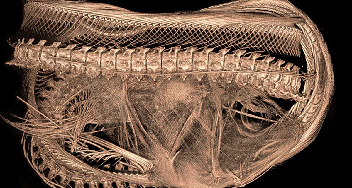
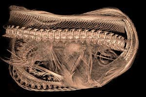
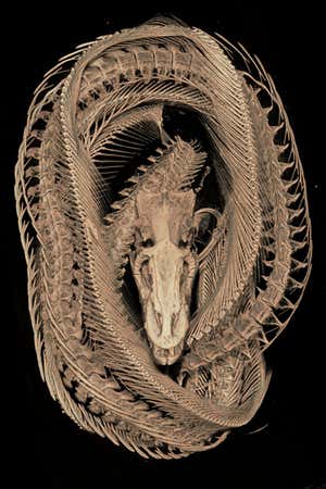
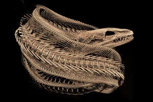
MORAY eels are known for their long, flexible spines and highly specialised double jaws that let them swallow large prey whole. Now, these unique skeletal features have been imaged in exquisite detail.
Karly Cohen at the University of Washington in Friday Harbor took these computerised tomography (CT) scans of two purplemouth moray eels (Gymnothorax vicinus) for her colleague Andrew Clark at the College of Charleston in South Carolina, who is studying their vertebrae. The images, of museum specimens, show the eels’ many vertebrae – 132 in total.
Karly Cohen
The scans also reveal their fearsome double jaws. These fish use one jaw to grab and hold their prey, and then their second jaw – which sits further back in their throat – shoots forward and drags the prey deep into the eel’s digestive tract. One of the eels still has a fish inside it that it ate whole. “The fish is way larger than the mouth of the moray eel itself, so this really shows you what they’re able to do with their cool morphology,” says Cohen.
She imaged these specimens, each around 80 centimetres long, by soaking them in ethanol to keep them moist, wrapping them in cheesecloth and curling them up tightly inside 3D-printed plastic cannisters to keep them in place.
Karly Cohen
Cohen’s work is part of a larger project aiming to perform CT scans of all 30,000 known species of fish, mostly using museum specimens. “CT is a non-destructive way to see inside fish and understand their skeletons, not just what they look like on the outside, so it’s a really valuable tool,” says Cohen.
[ad_2]
Source link