[ad_1]
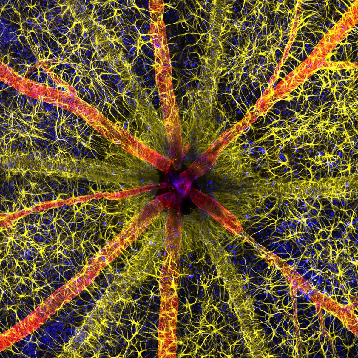
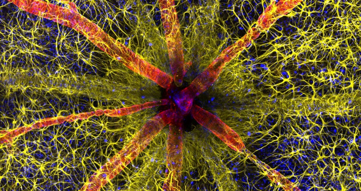
A rat’s optic nerve head
Hassanain Qambari & Jayden Dickson
THIS dazzling compilation of life under the microscope showcases some of the best photos entered into 2023’s Nikon Small World Photomicrography Competition.
Scooping the top prize is a picture of a rat’s optic nerve head (pictured above), which is where all the retinal nerve fibres that travel to the brain pass through before forming the optic nerve. The image is a powerful snapshot of the extreme intricacy of the eye’s retina. It was taken by Hassanain Qambari, assisted by Jayden Dickson, both of whom are at the Lions Eye Institute in Western Australia.
Qambari’s research focuses on detecting and finding treatments for diabetic retinopathy, in which high blood sugar levels cause blood vessels in the back of the eye to swell and leak, resulting in blurry vision and, sometimes, blindness.
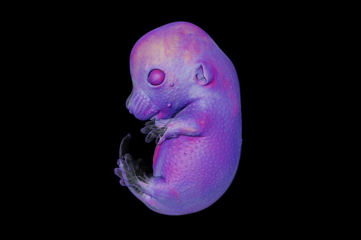
A mouse embryo
Dr. Grigorii Timin & Dr. Michel Milinkovitch
He said he entered the contest to showcase how complex this vascular network is. Locating the microscopic vessels was a “technically demanding challenge”, he added.
Another rodent features in the photo above – of a mouse embryo – taken by Grigorii Timin and Michel Milinkovitch at the University of Geneva, Switzerland. Achieving seventh place, the picture was captured using a technique called light sheet fluorescence microscopy.
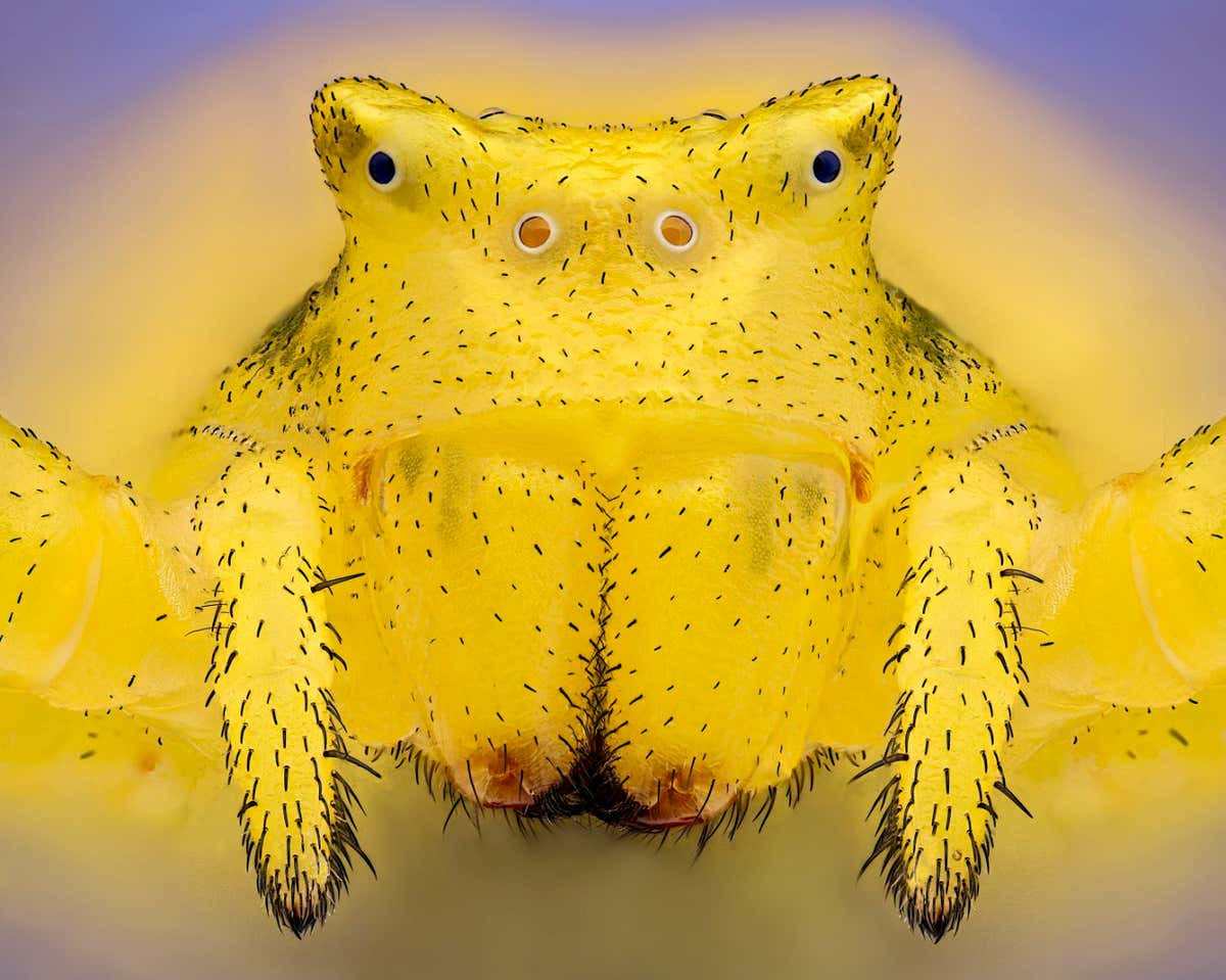
close-up of a crab spider
Sébastien Malo
Pictured above, winning an Image of Distinction award, is Sébastien Malo’s close-up of a crab spider (Thomisus onustus), the females of which can change colour between yellow, pink and white to blend in seamlessly with the flowers on which they both stalk prey and hide from predators.
Topics:
[ad_2]
Source link