
[ad_1]
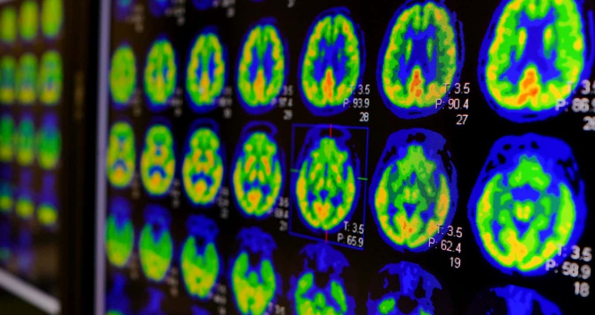
PET scans of a human brain
Utthapon wiratepsupon/Shutterstock
Researchers have drawn up the most detailed description ever created of the human brain, based on the thousands of different cell types that make it up.
The human brain cell atlas is a series of data sets about the various cell types within the brain and where they are located, defining this complex organ in more precise genetic and molecular detail than ever before.
The data comes from samples taken from deceased adults and children and from embryos and fetuses during pregnancy. The donors include some individuals who have certain medical conditions and others who don’t.
Samples from chimpanzees, gorillas and monkeys have also been included in order to shed light on brain evolution.
The resource has been compared to the enormous effort that went into the Human Genome Project, which led to multiple medical advances.
It is designed to lay the groundwork for further research into conditions that affect the brain, including neurological conditions such as Alzheimer’s and Parkinson’s disease, as well as mental health problems such as depression and schizophrenia. “If you’re going to explore a new country, you need a map so you know where you’re going,” says Sten Linnarsson at the Karolinska Institute in Stockholm, Sweden.
Previous attempts to visualise the brain have involved scanning techniques such as magnetic resonance imaging (MRI). While brain scanning can be invaluable for making medical diagnoses of individuals – for instance, by revealing a brain tumour – they typically can’t visualise anything smaller than about 1 millimetre.
The new atlas allows us to analyse the brain at a much smaller scale by identifying cell types taken from more than 100 different regions of the brain, depending on the species and age of the donors.
It was already known that there are many different types of brain cells, or neurons, partly based on the fact that they can look different or behave differently when grown in a dish. For instance, while most neurons send signals that trigger the next neuron down the line to fire, some send signals that stop the next neuron from firing.
But it was difficult to precisely identify the multitude of different neurons that exist until the advent of a technique called single-cell sequencing about 20 years ago.
This analyses the genetic material of a single cell to see which genes are turned on or off – in other words, which proteins it is making.
As single-cell sequencing techniques progressed, the US National of Institutes of Health began funding two initiatives, which coalesced into a set of 24 papers published on 12 October.
The first aimed to draw up a brain cell “census”, consisting of a detailed description of all the different cell types in the human brain, based on which of their genes are active. The second set out to identify exactly where in the brain those types of neuron are found in order to produce a cell-based atlas of the brain – although, at the moment, it exists only as a huge set of databases, rather than a single visual representation.
The first draft involved efforts by multiple international research teams, sequencing millions of brain cells altogether.
Linnarsson was involved in several of the projects, including carrying out single-cell sequencing of 105 anatomical locations from four adult brains. The project identified 3313 different types of brain cells.
Some insights into different medical conditions are already starting to emerge. “There are papers [due to come out] from geneticists that are mapping all the disease genes of brain diseases to our atlas,” says Linnarsson. “There will be insights there for sure.”
“There’s an enormous incentive for understanding brain function, not just because it’s such an intriguing question, but also because it can lead to therapies,” says Arnold Kriegstein at the University of California, San Francisco, who is part of the team drawing up atlases for the fetal brain at different stages of development.
“In future, researchers may be able to use these tools to study cell-specific and circuit-specific activity which underlies complex cognition and behaviour,” says Barbara Sahakian at the University of Cambridge, who wasn’t involved in the research. “This will not only advance our knowledge of the healthy brain, but may also lead to novel treatments for brain disorders.”
Topics:
[ad_2]
Source link