
[ad_1]

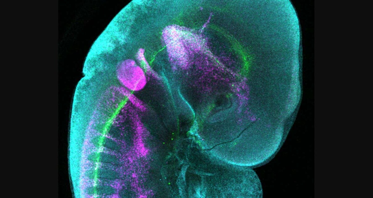
Chick embryo with fluorescent antibodies labelling parts of the developing nervous system
Erica Hutchins, UCSF
Scientists have visualised the early development of the nervous system in a chicken embryo.
Minyoung Kim at the University of California, San Francisco, dissected the 2-day-old embryo out of its egg and added fluorescent antibodies, which bind to specific proteins in the nervous system and make them visible.
An image of the embryo taken with a confocal laser scanning microscope shows its developing nerves in green.
The purple colour shows a group of cells called the neural crest that migrate through the embryo and form neurons in the gut, sensory nerves in the face and a diverse range of other cell types.
The cyan colour shows the presence of a protein called ELAVL1, which plays a role in neural crest development, says Erica Hutchins at the University of California, San Francisco, who oversaw the project.
“We use the chick embryo to investigate the molecular and cellular mechanisms of neural crest development because this model system develops similarly to human embryos, but develops outside the mother, allowing for easy manipulation of gene expression as well as live imaging approaches,” she says.
Hutchins and her team are investigating neural crest development in embryos because some congenital conditions are caused by the abnormal migration of these cells. These include Hirschsprung’s disease, in which nerves are missing from parts of the intestine, and familial dysautonomia, which can affect the ability to feel pain among other things.
Topics:
[ad_2]
Source link