
[ad_1]
The British Heart Foundation (BHF) has announced the winner of its annual Reflections of Research science image competition. This asks BHF-funded scientists to highlight their research into heart conditions by sharing pictures that were taken as part of their work.
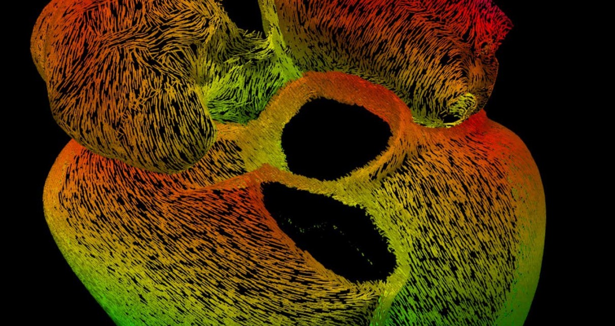
“Paths of the heart”, which maps the thousands of muscle cells that make up the wall of the organ, won top prize at the competition
Marina Strocchi/King's College London/British Heart Foundation
First prize went to this computer-generated image that maps the thousands of muscle cells that make up the wall of the heart. Each tiny line represents a bundle of cells that sends electrical signals to each other.
The “Paths of the heart” image was submitted by Marina Strocchi at King’s College London, who hopes that by visualising how electrical signals travel through the heart we can better predict how individuals will respond to different treatments for cardiovascular conditions.
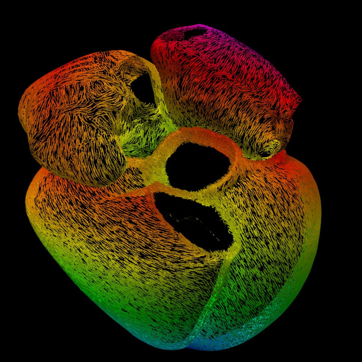
“Seeing through the heart” highlights the blood vessels around the outside of the organ and the electrical system at its centre
Judy Sayers, University of Oxford, British Heart Foundation – Reflections of Research
Captured by Judy Sayers at the University of Oxford, “Seeing through the heart” won the supporters’ favourite category at the competition. The glows of orange depict the blood vessels around the heart, with the thick branch on the left being the coronary artery, while the largest bright spots show the upper chambers of the heart. Nestled in the middle of the organ is its intertwining electrical system, which causes the heart to beat.
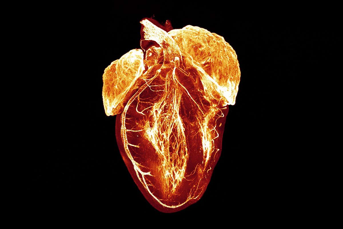
“A close-up view of vascular first aid kits” shows a blood vessel in minute detail
Sammy El-Mansi/Queen Mary University of London/British Heart Foundation
One of several shortlisted entries, “A close-up view of vascular first aid kits” was submitted by Sammy El-Mansi at Queen Mary University of London and zooms in on a blood vessel. The specks of green, red and yellow are stores of a molecule called von Willebrand factor, which the vessels’ cells release to create blood clots. The splashes of blue denote the genetic information of each cell and the slivers of white are endothelial cells, which form the lining of the blood vessels.
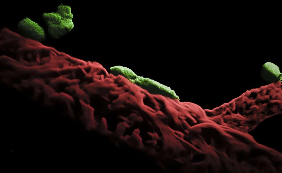
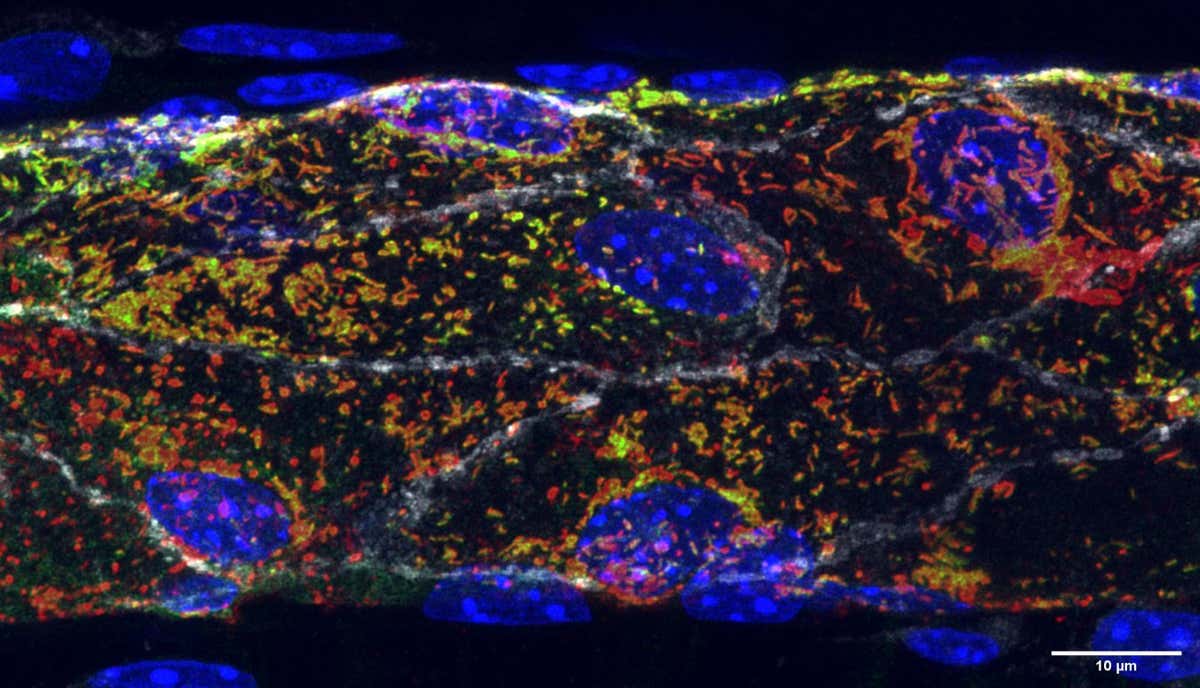
“The radiant microvascular realm” shows the interaction between blood vessels and immune cells
Dr Loïc Rolas/Queen Mary University of London/British Heart Foundation
An image from Loïc Rolas, also at Queen Mary University of London, looks at the outside of a mouse’s blood vessel, represented by the deep red structure at the bottom of the image. The green bodies on top of the blood vessel are mast cells – immune cells that control blood flow and inflammation.

“A window into the heart of artificial intelligence” was created by an AI generator but has some structural flaws
Michelle Williams/University of Edinburgh/British Heart Foundation
Unlike the previous entries, which were created from laboratory studies, this vibrant image was the product of the AI image generator DALL-E.
Michelle Williams at the University of Edinburgh, UK, prompted the generator to create “an image of the heart from a computed tomography scan, digital art”. As striking as the result is, there are a few anatomical errors, such as the structure and pattern of the blood vessels.
Williams, along with her team, is investigating whether healthcare professionals can distinguish between real and AI-generated heart scans.
Topics:
[ad_2]
Source link